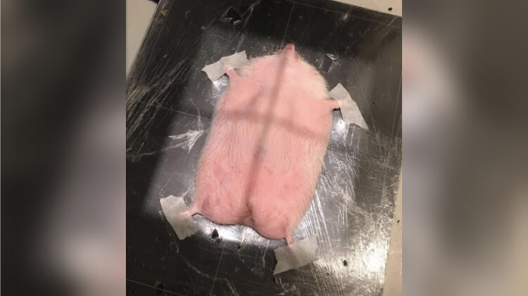
Believe it or not, thousands of hedgehogs come through the doors of veterinary clinics every year, and around two thirds of them will require an X-ray. These small, prickly creatures are a popular choice of pet for many animal lovers, but their unique physiology and natural instincts make them particularly prone to injury and illness. X-rays are an important diagnostic tool in the treatment of these animals, helping veterinarians to identify a range of health issues.
But how exactly is an X-ray performed on a hedgehog? First, the hedgehog is carefully placed under anesthesia to keep them calm and still during the procedure. Next, a small plate is placed behind the area of interest, and an X-ray beam is directed at the plate. The beam passes through the hedgehog’s body, and the resulting image is captured on the plate. The plate is then removed and the image is processed and analyzed by the veterinarian.

While X-rays can help detect a range of health issues, they are particularly useful in identifying broken bones, foreign objects, and other internal injuries that may not be visible from the outside. They can also be used to monitor the progress of treatment or recovery in hedgehogs with chronic health conditions.
While hedgehogs may not be the first animal that comes to mind when you think of X-rays, they are a common and important patient for veterinary clinics around the world. As with any pet, it’s important to take proper care of hedgehogs and seek medical attention when needed. And for these little creatures, that often means undergoing an X-ray to help keep them healthy and happy.

Leave a Reply