
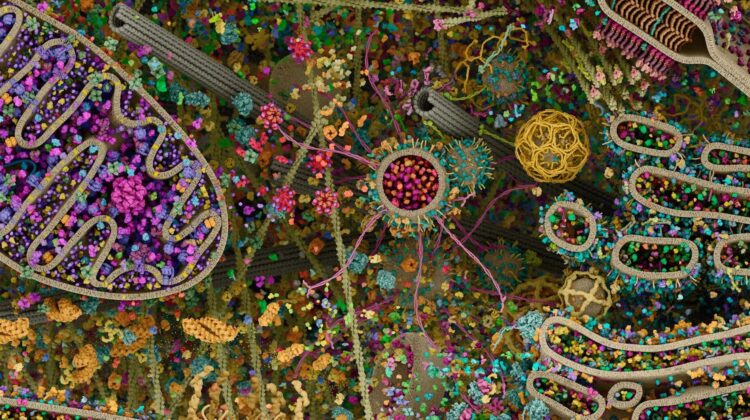
A remarkable scientific visualization is offering people an unprecedented look inside the microscopic world of the human cell — revealing a level of complexity that many have described as an entire universe hidden within the body.
Built using real molecular and structural data gathered through X-ray crystallography, electron microscopy and advanced computational modeling, the reconstruction transforms scientific measurements into an immersive three-dimensional scene.
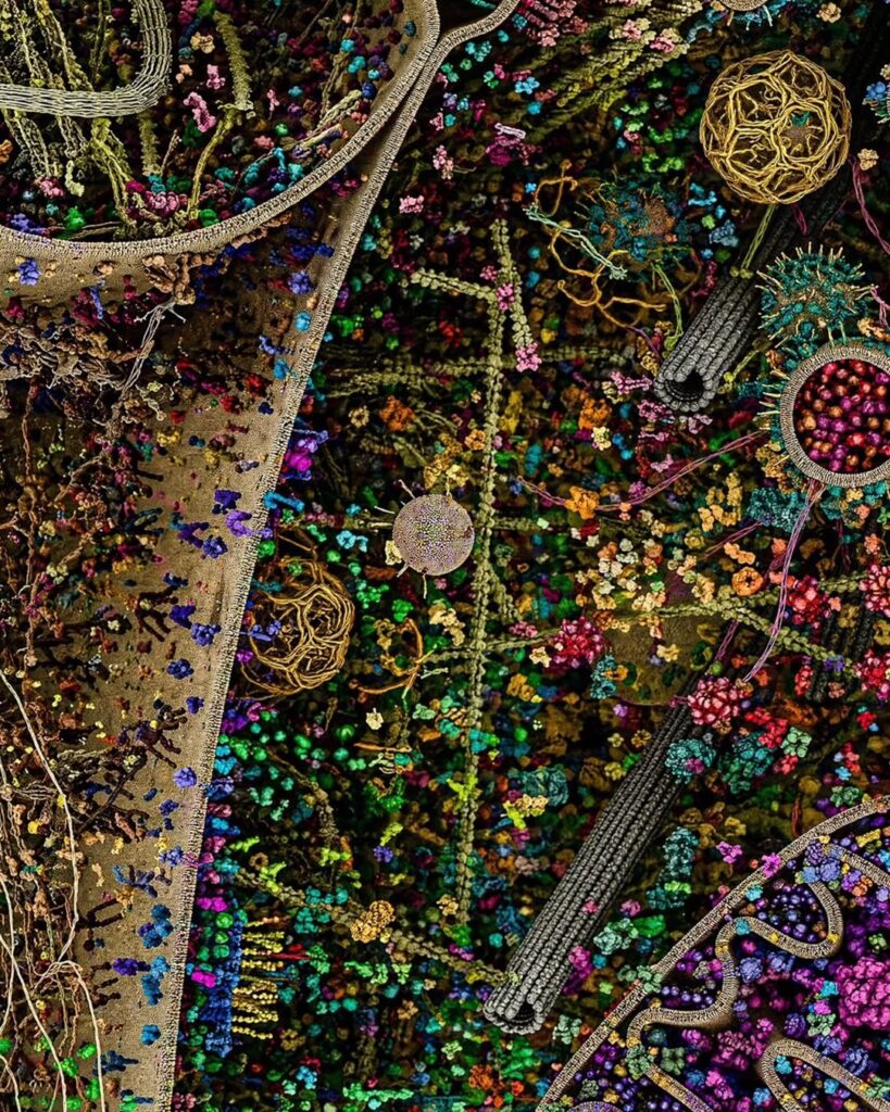
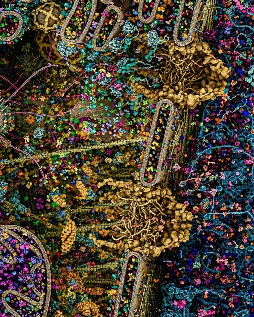
Rather than presenting the cell as a simple diagram, the project allows viewers to see its dense internal environment in extraordinary detail — from protein structures and membranes to the countless molecular interactions that keep life functioning.
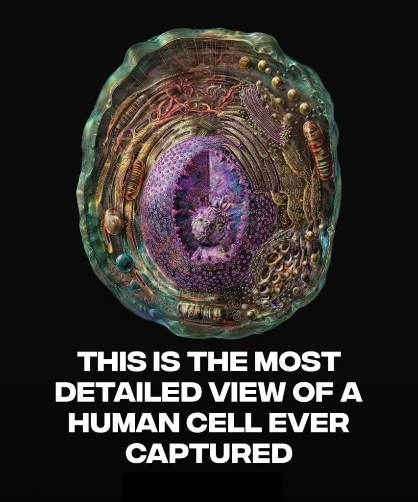
The transparency and lighting used in the visualization create the impression of looking directly inside a living system, helping translate highly technical biological data into something both scientifically meaningful and visually accessible.
Researchers and science communicators say such reconstructions are becoming increasingly valuable because they make it easier for both specialists and the public to understand how cells actually work beyond textbook illustrations.
Human cells are extraordinarily busy environments, packed with microscopic machinery responsible for producing energy, transporting materials, processing information and maintaining survival.
By combining multiple scientific imaging methods, these reconstructions can provide more realistic educational tools for biology, medicine and biotechnology.
For many viewers, the image serves as a striking reminder that even the smallest components of the human body contain staggering levels of organization and sophistication.
As imaging technologies continue to improve, scientists are expected to create even more precise visual models, potentially improving medical education, pharmaceutical research and our broader understanding of disease at the cellular level.
What appears invisible to the naked eye is, in reality, a vast biological system operating continuously inside every person.
Why detailed cell imaging matters for science and medicine
Advanced cell visualization is not only reshaping biology education — it may also play an important role in future healthcare innovation. Better molecular imaging can support disease research, drug development, cancer studies and personalized medicine by helping scientists understand how cellular structures behave under different conditions. As medical technology evolves, breakthroughs in microscopic imaging could influence everything from diagnostics to biotechnology investment.
Leave a Reply