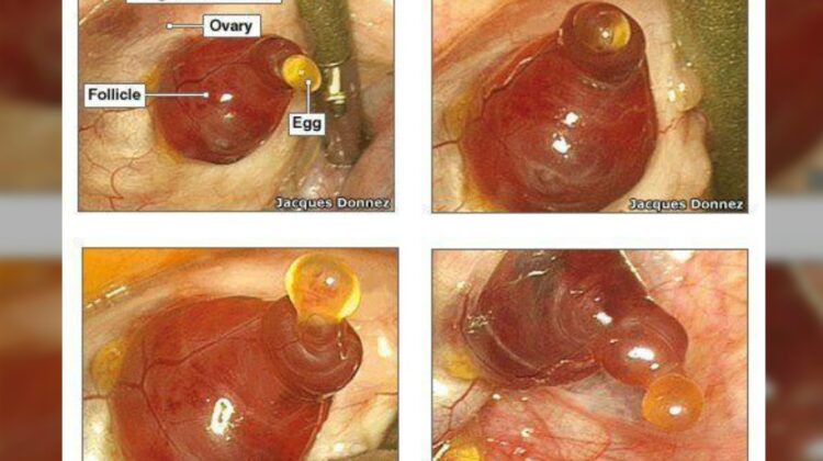
In a groundbreaking moment for reproductive science, Dr. Jacques Donnez, the head of gynecology at UCL in Brussels, Belgium, has achieved a remarkable feat—photographing the elusive event of human ovulation for the first time in history. The significance of these images lies not only in their rarity but in the valuable insights they provide into the intricacies of ovulation, a phenomenon occurring approximately 13 times a year in the average American woman.
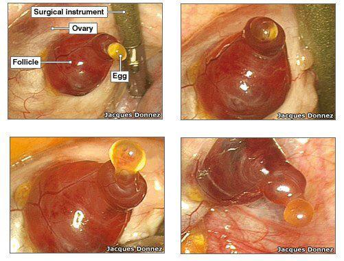
Ovulation, a swift process lasting a maximum of 15 minutes, has long eluded clear documentation due to its infrequency and unpredictability. Dr. Donnez stumbled upon this remarkable opportunity while preparing for a partial hysterectomy on his 45-year-old patient. The spontaneous occurrence of ovulation during this procedure allowed him to capture, in exquisite detail, the release of a mature egg from the ovary—an event crucial to human reproduction.
Published in the esteemed professional journal Fertility and Sterility, these images challenge prior beliefs about the rapid explosiveness of ovulation. Contrary to previous assumptions, Dr. Donnez’s series of photographs reveal a more gradual process, unfolding over nearly 15 minutes. “The release of the oocyte from the ovary is a crucial event in human reproduction,” notes Dr. Donnez. “These pictures are clearly important to better understand the mechanism.”
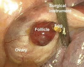
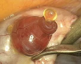
Dr. Alan McNeilly from the Medical Research Council’s Human Reproduction Unit in Edinburgh, UK, emphasizes the historical significance of these images. Until now, clear documentation of human ovulation had been limited to fuzzy images, with successful captures only seen in other animal species. Dr. McNeilly underscores the rarity of witnessing ovulation in real-time, describing it as “a pivotal moment in the whole process, the beginnings of life in a way.”

The images provide a detailed view of the ovulation process. Initially, the mature follicle, a fluid-filled sac on the ovary’s surface containing the ovum (egg), is observed. Enzymes then break down the follicle’s tissue, leading to the emergence of the ovum. A red-colored ballooning and a minuscule hole appear at the top of the follicle, signaling the ovum’s release. Protected by a sac of support cells, the ovum embarks on its journey through the fallopian tube into the uterus.

Following ovulation, there is a brief window of approximately 24 hours during which the ovum remains viable for fertilization. Should live sperm be present at the cervix or in the uterus before ovulation, conception could occur without consecutive sperm introduction. Sperm typically retain viability for about 72 hours (3 days) within a woman’s body.
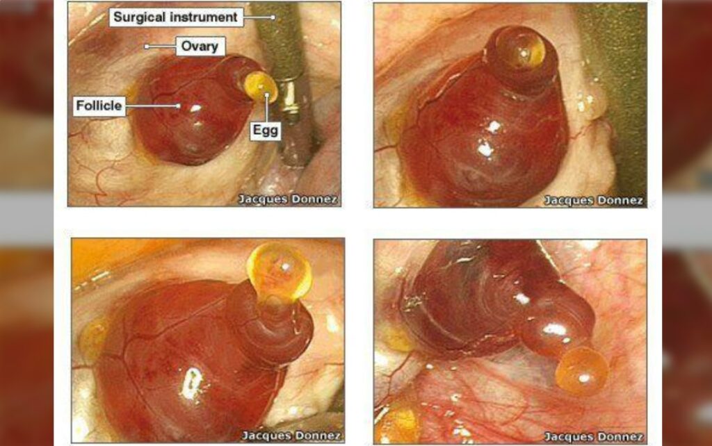
These historic photographs not only mark a significant milestone in reproductive science but open new avenues for understanding the complexities of human ovulation, shedding light on a process that has remained largely enigmatic throughout history.

Leave a Reply