
Unusual and unsettling images circulating online have sparked curiosity about how medical scans work during pregnancy — especially when it comes to MRI technology.
While these images may look alarming at first glance, they actually reveal something important about how doctors study fetal development and why certain imaging methods are used more cautiously.
What the Images Show
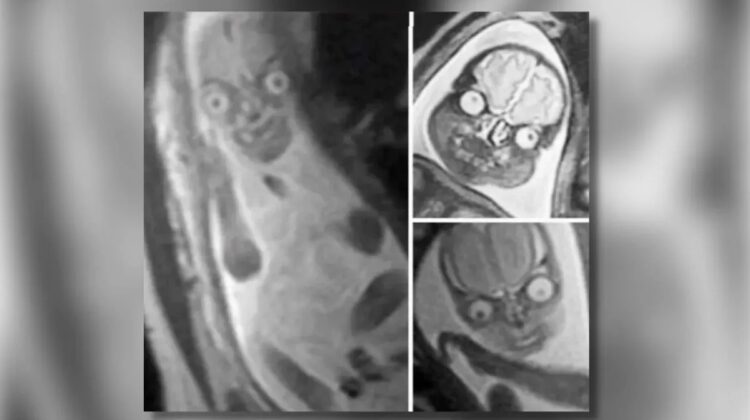
Some widely shared images appear strange or even frightening, showing a fetus in a way that looks very different from typical ultrasound pictures.
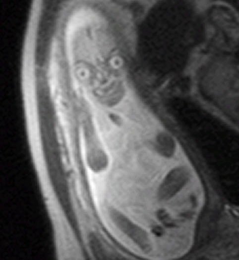
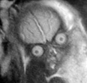
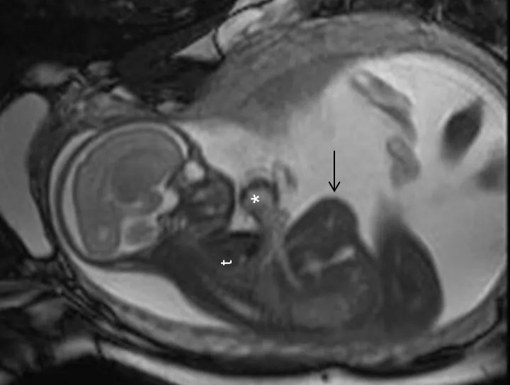
Unlike the soft, familiar outlines seen in ultrasounds, MRI scans can produce highly detailed images of internal structures. This includes the brain, eyes, and other soft tissues, which can appear exaggerated or distorted due to how the technology works.
These images are real and have been used in scientific research to better understand fetal development.
How MRI Scans Work
Magnetic Resonance Imaging (MRI) uses powerful magnetic fields and radio waves to create detailed images of the inside of the body.
Because the human body is largely made up of water, MRI scans are especially effective at capturing differences between soft tissues. This allows doctors to distinguish between areas like:
- The brain
- Eyes
- Facial structures
- Internal organs
The result is a highly detailed image — sometimes far more detailed than what other imaging methods can provide.

Why the Images Look So Unusual
MRI images can look unfamiliar because they highlight contrasts in soft tissue rather than providing a simple outer shape.
Differences in how tissues respond to magnetic signals can make certain areas appear brighter or darker. This can create unusual visual effects, especially when viewing a developing fetus.
While these images may seem unsettling, they are simply a result of advanced imaging technology revealing layers of the body that are not normally visible.
Why MRIs Aren’t Common in Pregnancy
Despite their accuracy, MRI scans are not routinely used during pregnancy.
In most cases, doctors rely on ultrasound imaging, which is safe, widely available, and effective for monitoring fetal development.
MRI scans are typically only recommended when:
- Ultrasound results are unclear
- Doctors need more detailed information
- There is a concern about specific developmental issues
When used, MRIs are generally performed during the second or third trimester, when necessary.
Safety Considerations
MRI technology does not use radiation like X-rays or CT scans. However, because it involves strong magnetic fields, doctors take a cautious approach when considering its use during pregnancy.

Medical professionals weigh the benefits against the need for additional imaging before recommending an MRI.
A Tool for Advanced Diagnosis
In certain situations, MRI scans can provide critical insights that other imaging methods cannot. They are particularly useful for examining the fetal brain and detecting conditions that may not be visible through ultrasound alone.
Advances in imaging technology have made MRI scans more effective and safer, allowing doctors to make better-informed decisions when monitoring pregnancy.
Understanding the Bigger Picture
While the images may appear unusual or even unsettling, they represent the incredible ability of modern medicine to look inside the human body in ways that were once impossible.
Rather than something to fear, these scans highlight how technology continues to improve our understanding of human development and health.
Modern healthcare relies heavily on advanced diagnostic tools such as MRI imaging systems, ultrasound technology, prenatal screening services, medical imaging software, and hospital diagnostic equipment. These technologies play a vital role in early detection, accurate diagnosis, and improved patient care, helping medical professionals monitor complex conditions and ensure better health outcomes for both mothers and babies.
Leave a Reply